Image

3DHEALS Legal Conference- August

3D Printing Poses Unique Security Risks for Medical Devices

Want to write a piece for 3DHEALS Expert Corner? Email us: info@3dheals.com

3DHEALS Legal Conference- August

Although cyber threats are nothing new to the healthcare industry, the rise of 3D printing in the medical industry presents unique cybersecurity challenges for medical device manufacturers and hospitals.  In July 2016, researchers at the New York University (NYU) Tandon School of Engineering found that 3D printing poses cybersecurity risks in the manufacturing process that could affect the reliability of the end product.  The study found that a hacker could potentially alter the printer head orientation without detection, affecting the strength of the device by as much as 25 percent.  Alternatively, a hacker could manipulate a 3D printer while it is connected to the internet to introduce internal defects as the device is being printed.

Although 3D printing offers many manufacturing benefits by making it possible to print customized devices on demand offering better fit and greater comfort for patients, the digital transmission of design files makes it more challenging to ensure sensitive confidential patient information is protected.  Confidential patient information contained within patient imaging  (e.g., computed tomography (CT) or magnetic resonance (MR) imaging) is vulnerable to being compromised or stolen in the digital transmission process.  Mishandling HIPAA-protected information carries heavy penalties and has resulted in more than $9 million in fines in 2016, according to the U.S. Department of Health & Human Services.

Legal Issues of Healthcare 3D printing

The FDA has acknowledged the need for effective cybersecurity measures to ensure the safety and efficacy of medical devices and protect patient health.   In October 2014, the FDA issued a guidance entitled Content of Premarket Submissions for Management of Cybersecurity in Medical Devices. The guidance, although not specific to 3D printed devices, sets forth recommendations for manufacturers to develop cybersecurity controls during the design and development stages of a medical device as well as in preparing FDA premarket submissions for software and devices using software.  The FDA urges manufacturers to establish a cybersecurity vulnerability and management approach as part of the software validation and risk analysis required by 21 C.F.R. part 820.30(g) governing design validation.  The FDA also recommends that device manufacturers adopt security measures to protect medical devices including, but not limited to, limiting access to devices to trusted users only and ensuring trusted content.

While the FDA recommends the use of design controls to assure medical devices will maintain their integrity from the point of origin to the point at which the device leaves the control of the manufacturer, 3D printing raises the question, “When is a device considered to have a left a manufacturer’s control?”  Unlike traditionally-manufactured medical devices that get physically shipped, 3D printed medical devices are sent electronically in the form of a CAD file from the manufacturer often to a service bureau to print off-site.  Has the device left the manufacturer’s control after the CAD file was sent?  After the medical device is 3D printed?   The law is unsettled on these issues.

The FDA has also addressed the postmarket management of cybersecurity issues in medical devices.  In January 2016, the FDA released a draft guidance entitled Postmarket Management of Cybersecurity in Medical Devices, which sets forth the FDA’s post-market recommendations.  The FDA encourages manufacturers to monitor, identify, and address cybersecurity vulnerabilities as part of their postmarket management of medical devices and sets forth elements of an effective postmarket cybersecurity program.  The program includes defining the risk acceptance criteria of a device; identifying cybersecurity signals and creating a process for intake and handling of vulnerability information; conducting cybersecurity risk analyses; and implementing device-based features to mitigate the risks on a device’s functionality.

In May 2016, the FDA issued a draft guidance regarding 3D printed devices for the first time.  The guidance entitled Technical Considerations for Additive Manufactured Devices addressed technical considerations associated with the design, manufacturing, and device testing of 3D printed devices.  Although not addressing cybersecurity, the FDA acknowledged patient safety risks associated with the file format conversion process.  Because additive manufacturing requires files to be compatible across the various software applications that are used, the file formats must be converted to ensure compatibility and this process creates a risk of errors that can negatively affect the shape and dimensions of the finished device.  The FDA explained that “patient-matched devices that follow the patient anatomy precisely are especially vulnerable” to errors in the file conversion process because “anatomic curves are typically geometrically or mathematically complex and can create difficulties when calculating conversions.”  The FDA recommends that manufacturers “test all file conversion steps with simulated worse-case scenarios to ensure expected performance, especially for patient-matched devices” and maintain and archive final device files in standardized formats that are able to house additive manufacturing information.

It remains to be seen whether the FDA will issue a guidance regarding cybersecurity considerations specific to 3D printed devices.  However, device manufacturers and hospitals should regularly conduct cybersecurity risk assessments in conjunction with their Information Technology, risk management, and legal departments under the protection of applicable legal privileges to minimize exposure to regulatory or legal action.

tabibkhoei,f_hi-c

Farah Tabibkhoei is a member of the Complex Litigation Group at Reed Smith LLP.  Her practice focuses on medical device product liability, managed care disputes, and 3D printing.  Farah can be reached at FTabibkhoei@ReedSmith.com.

You may also like the following articles:

3D Printed Orthopedic Implants in China and the Challenges in Commercialization

Government Shutdown and 3DHEALS: There is a Connection, Believe or Not
Decentralized Healthcare — Part II: Breaking It All Down

copy right Stanford Healthcare

Segmentation: The Real Struggles Behind Converting DICOM to Patient-specific 3D Printable Models

Want to write a piece for 3DHEALS Expert Corner? Email us: info@3dheals.com

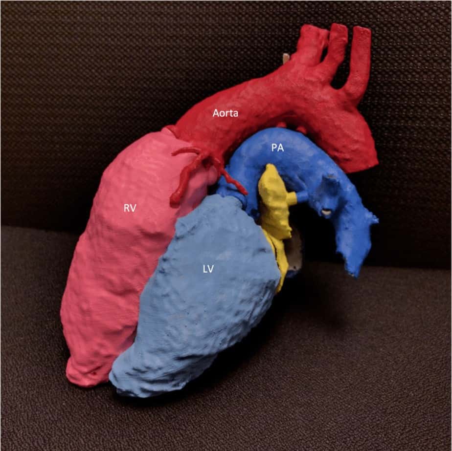
Disarticulating Congenital Heart - FDMAnatomy: Disarticulating Congenital Heart Disease ; Purpose: Provide example of Transposition of Great Arteries Mustard Switch procedure; Print Technique: Fused Deposition Modeling (FDM); Image Source: Computed Tomography of the Chest, 1 mm voxel resolution; Segmentation Difficulty: Very Difficult; ensuring no overlap of structures was difficult, many different models; Credit: Chris Letrong, Shannon Walters — Stanford University Department of Radiology, 3D and Quantitative Imaging Laboratory.

In a future healthcare world, physicians may be able to 3D Print any particular part of a patient’s anatomy with the press of a button. At present, however, precision DICOM image segmentation is more complex than many articles and presentations seem to suggest. I use medical 3D software daily to accomplish 3D replication, visualization, and quantification. Even before 3D Printing, I observed that most automatic and manual segmentation tools could use significant improvement. Since attempting more than 50 patient-specific 3D anatomic models, this observation is reinforced. Perhaps the repeatability and usability of many segmentation tools suffer due to lack of user input during the development of algorithms. Medical 3D software developers may have an opportunity to improve segmentation algorithms by leveraging user knowledge and preferences.
I perceive a lack of connection between those who spend thousands of hours using segmentation and those who design software that segments DICOM data. Many anatomic structures that may need 3D printing (and thus, segmentation) are not clearly delineated, homogenous, isolated, and uniform due to pathology or anomaly. Additionally, image quality adds the variables of graininess, artifact, slice thickness, and anatomic coverage. These variations in quality image data from CT or MR scanners increase the difficulty to successfully implement automatic segmentation. Perhaps we can move toward semi-automatic segmentation, with software vendors accepting various logic to improve segmentation time and accuracy. Other limitations for segmentation exist such as user familiarity with software tools, understanding of anatomy, and understanding the need for a 3D printed model. This post will focus on segmentation tools, provide a perspective on current limitations based on image quality, and propose some actions to help us arrive at the distant future of truly automatic segmentation.

What is segmentation?

Each vendor has a unique set of terminology for this, but the essence of segmentation is to identify and isolate voxels that represent any anatomy of interest. Two implementations of segmentation are; a) assigning a mask to a dataset indicating active voxels or b) deletion/removal of voxels not included in segmentation. Managing the models and masks is also unique in methodology and terminology per software vendor.
Methods of segmentation are both automatic and manual. Automatic segmentation can be threshold- or atlas-based. Threshold-based segmentation uses pixel brightness and patterns throughout the DICOM data to isolate or remove structures.  Atlas-based segmentation uses a database of anatomic structure shapes and attempts to find similar patterns in the current DICOM dataset. Many vendors have a threshold-based automatic segmentation method and “freehand” manual segmentation method.

What should be segmented?

The segmentation in this post refers to identifying and isolating anatomic structures within DICOM datasets. Structures are typically differentiated in the datasets by either discreet pixel intensity values ore relational differences in signal intensity. The context of any given DICOM acquisition must be taken into account to understand which intensity values represent which anatomic structures; contrast, dose, timing, and patient status can impact the pixel intensity of any given organ.
For CT Scans, brightness measures are standard for various structures across most scanners; but many factors can affect whether the image accurately reflects such brightness with the correct patterns. The CT brightness measure is referred to as Hounsfield Units (HU). MRI signal intensity is dependent upon habitus, coil selection, magnetic fields, distance to coil, and much more. With more variables, MRI has a higher possibility of signal variations when multiple factors contribute. The signal intensity using phased-array MRI coils can cause gradients of signal intensity for a structure, such as the posterior surface of a kidney measuring double the signal intensity of the anterior surface.
Ultimately, segmentation of any anatomic structure is largely based on identifying the voxel intensity values which represent it. This is likely why most automatic segmentation tools are based on threshold. Unfortunately, many factors hinder optimal imaging that make such segmentation a simple task.  

Issues with segmentation

Several issues with threshold-based segmentation are;
Heterogeneous Structures: osteoporosis is an example; a patchy-looking bony structure rather than a nicely delineated bone shape of a normal young person.
Image Noise: mottles the appearance of the entire dataset, making homogenous structures appear heterogeneous. This will impact the ability to identify in entire organs and/or shapes.  
Artifacts: metal implants, various types of motion, and other artifacts contribute to inaccurate representations of anatomic structures. At present, I have not witnessed any threshold-based segmentations able to correct for artifacts.
Several issues with atlas-based segmentation are:
Non-standard anatomic representations: Many 3D Prints are likely to be of non-standard anatomy; atlas databases are typically built upon normal anatomy.
Image Noise: mottles the appearance of the entire dataset, making border detection much harder for the algorithms to identify.
Artifacts: metal implants, various types of motion, and other artifacts contribute to inaccurate representations of anatomic structures. Borders of affected structures will not conform to atlas models because the signal characteristics will not match any in the database.  

What can be done?

First and foremost, all software must keep a robust manual segmentation tool as a backup to any automatic/semi-automatic method. Despite any adherence to my suggestions or others’, it is unlikely in the near-term that every patient condition and image type can be accommodated using automatic segmentation. With that said, I believe that three steps can help vendors achieve greater results in providing automatic segmentation that works despite the many image quality issues that arise.

  1. User-driven development of segmentation algorithms
  2. Semi-automatic approach, allowing logic to drive the segmentation approach
  3. Validation of segmentation algorithms on standardized datasets

1. User-Driven Development

Developers should request feedback/involvement from users to improve segmentation algorithms. Much of current medical 3D software is likely designed around radiologist workflows, largely due to radiologists being the most obvious users of 3D software and having a role in purchasing decisions. There is a growing cohort of 3D Imaging Laboratories that utilize non-radiologists (technologists and others) to perform advanced functions on patient DICOM datasets. This non-radiologist population will likely grow as 3D Printing and other kinds of visualization and quantification proliferate. Vendors that singularly accommodate radiologists concerns may not achieve the needs of other users.

2. Semi-Automatic Segmentation

Given the myriad of image quality issues that will not dissipate soon, developers should acknowledge the need to overcome issues of heterogeneity, artifact, and image noise. Perhaps this could manifest as a questionnaire that optionally appears when segmentation begins. This questionnaire may ask the user about image quality factors; whether and where artifacts exist, ask users to set bounding boxes, and perhaps ask the users to quickly identify each structure in the dataset with a click. Using such logic, perhaps future segmentation can use atlas or threshold tools better to identify the desired anatomy with much more information from which to base segmentation algorithms on.

3. Validation of Segmentation

This may be far-fetched, but it would be nice to have an independent set of DICOM data from which 3D software can be applied to and potentially scored. Segmentation could be a single category of analyses, with subcategories of the vendor, MR, CT, and even further subcategories of an artifact, image noise, and so on. If all developers were forced to test on the same data, users could more effectively evaluate which tools might be best for their specific location and requirements. To fairly apply this tool, anonymized data from each CT/MR vendor with all the different kinds of equipment and image quality variable representations must be collected and prepared for analysis.

Where to begin?

Similar to gaining traction from 3D Printer vendors toward medical community needs, our community will need to show the returns on vendors investing resources to solve our problems. As this community grows, the issues will become more important. Radiologists involved with 3D printing at this point are in a position of leverage and should begin demanding that segmentation tools accommodate the myriad of needs that 3D printing will ultimately present. Perhaps the suggestion of a semi-automated approach is something that will benefit radiologists in other workflows than 3D Printing. All of us can track issues, articulate them carefully and think about ways to rate software based on objective measures such as the validation method suggested above. Lastly, without direct involvement with the developers, it is difficult to perceive how segmentation tools will ever truly meet the needs of the users, their needs, and the data they are forced to work with.
Shannon Walters, MS RT(MR), Stanford University Department of Radiology, 3D and Quantitative Imaging Laboratory
Shannon Walters, MS RT(MR), Stanford University Department of Radiology, 3D and Quantitative Imaging Laboratory  
Shannon been a radiologic technologist since 1998 and completed a Masters of Information Systems in 2014.  He has worked in Stanford 3D and Quantitative Imaging Laboratory since 2008, assuming the role of Manager in 2013. The field of advanced visualization is a perfect fit for Shannon’s intense interests in computers and healthcare.  Shannon has been involved with 3D Printing since 2013 and has generated more than 50 patient-specific models as of mid-2016.

Related Articles:

Part 1: Considerations for Implementing a 3D Printing Core Service in Your Hospital: A Technical Analysis

Part 2: Considerations for Implementing a 3D Printing Core Service in Your Hospital: A Technical Analysis

Interview: Jeffrey Sorenson, President and Chief Executive Officer of TeraRecon

blank

Infographics: Legal Landscape of Healthcare 3D Printing Innovations

Legal Issues of Healthcare 3D printing

Infographics: Legal Landscape of Healthcare 3D Printing Innovations

Healthcare industry is notoriously highly regulated. The legal issues surrounding healthcare 3D printing technologies are not only complex but often completely unknown to both the scientific and the entrepreneur communities. As the healthcare 3D printing community grows exponentially, even the legal experts themselves are sometimes struggling to keep up with new problems facing with the rapidly growing field and innovations. Come and join us at 3DHEALS August event focusing on some of the most interesting legal topics surrounding healthcare 3D Printing.

3D printed heart

Legal: Take Care Not to Trigger HIPAA When Outsourcing Medical 3D Printing

Want to write a piece for 3DHEALS Expert Corner? Email us: info@3dheals.com

3D printed heart

3D printed heart -copyright Materialise

Written by Erik Birkeneder and Valerie Breslin Montague
Doctors are increasingly printing 3D models of a patient’s anatomy to plan for surgery or to aid in diagnosis.  For instance, a cardiac surgeon may take an MRI or CT scan of a patient’s heart to create a plastic model of the heart to plan a valve repair. However, many hospitals do not have 3D printing equipment in-house, so the hospital may send the MRI or CT scans to a company that specializes in 3D printing. Sending these scans outside the walls of the hospital could trigger the privacy and security requirements of HIPAA.

When health care providers send data with certain patient “identifiers” to third parties, the arrangement generally triggers HIPAA. In the case of 3D printing, the substance of what is transmitted, oftentimes a scan of an organ or body part, may not be deemed to be “identifiable” under HIPAA. For example, patient identifiers include the obvious information like names and social security numbers, but also include “biometric identifiers, including finger and voice prints,” “full face photographic images and any comparable images,” and “any other unique identifying number, characteristic or code.” Although many organ, tissue, bone and other body part scans likely will not be deemed to identify a particular patient, others might, such as images that contain fingerprints or dental models.

Prior to transmitting any images or data, health care providers must analyze whether the information to be sent to the 3D printing company is subject to HIPAA (or any other state or federal laws protecting patient confidentiality). If the information does contain patient identifiable information, referred to as “protected health information” under HIPAA, either due to the content of the image, the patient’s name or record number on the image or other data identifying the patient in what is transmitted, the provider must enter into a HIPAA business associate agreement with the 3D printing company.

3D printing companies also should take care when preparing models for hospitals, physicians and other health care providers. Generally, if a 3D printing company is the recipient of protected health information, the company becomes a “business associates”, subject to a number of the HIPAA regulations, such as requirements to adopt designated policies and procedures, conduct a security risk assessment and train the company’s workforce on HIPAA compliance. The company can avoid these compliance efforts by working with its health care clients to ensure that no protected health information is transmitted during the arrangement.

Disclaimer: The foregoing is not intended to convey or constitute legal advice, and is not a substitute for obtaining legal advice from a qualified attorney. You should not act upon any such information without first seeking qualified professional counsel on your specific matter.
JOIN US FOR A MORE IN-DEPTH DISCUSSION ON THE LEGAL ISSUES RELEVANT TO HEALTHCARE 3D-PRINTING ON AUGUST 10TH, 2016
thumb_CHJ_8593_1024

Authors:
Birkeneder
Erik Birkeneder is an intellectual property attorney at Nixon Peabody that focuses on health care related patents. Erik also serves as outside general counsel for a number of digital health companies and helps them navigate the unique privacy, and other regulatory hurdles that are facing this industry, including in 3D printing. He has a Master’s in Biomedical Engineering from University of Wisconsin Madison where he performed research on the impact of neuropeptides on wound healing in diabetics, and has a law degree from University of Minnesota.

https://angel.co/erik-birkeneder
linkedin
Valerie Montague Breslin
Valerie Montague represents a variety of health care providers, digital health vendors, senior living facilities, nonprofit trade associations, life sciences companies and vendors of health care providers. Valerie is a Certified Information Privacy Professional/United States (CIPP/US), the preeminent credential in the field of privacy.

3D Printing Heart

Part 2: Considerations for Implementing a 3D Printing Core Service in Your Hospital: A Technical Analysis

Want to write a piece for 3DHEALS Expert Corner? Email us: info@3dheals.com

(Cont’d) Considerations for Implementing a 3D Printing Core Service in Your Hospital: A Technical Analysis – Part 1

Verification and Labeling

After completing your segmentation and modeling work, you may be in a hurry to get your part on the 3D printer. However, there is an opportunity to unintentionally introduce errors in the above steps. Prior to printing, the accuracy of your final file should be verified against the original DICOM imaging. Did you take liberties and over-smooth or remove a key feature from the model? Did you cut away a structure that would be an important landmark for the surgeon? I would highly recommend a software solution that allows the user to overlay the STL surfaces back on the Dicom data. This will allow you to verify accuracy (and establish credibility with your surgical colleagues) as well as make subtle adjustments or refinements to the model.

Contour verification of prepared heart

Figure 4- Contour verification of prepared heart model demonstrated in Mimics Innovation Suite

Ensuring traceability of your 3D prints will also reduce the opportunity for making errors with your 3D printing program. As you scale your operation and build greater volumes of models, it is critical to understand what models are coming off the printer and what case they each belong to. To reduce the chance of mixing models or providing the wrong model to a surgeon, each anatomical model should be pre-labeled with software prior to printing. Use a requisition number that will trace back to the medical records to ensure traceability of your 3D models. You will also want to use labels if you create mirror images or want to clearly indicate what side of the patient the model was derived from. This will reduce the chance for operator confusion and eliminate the chance they may operate on the wrong side of the body.

figure 5

Figure 5- Applying a text label to a heart model using Mimics Innovation Suite

Communication

Close collaboration between personnel is key in this process. It is necessary to define the scope and use of the model with the surgeon or interventionist prior to starting the process in addition to verification near the end. This can be facilitated through web meetings or face-to-face discussion. It can also help to have a software solution capable of exporting a file format which can be interrogated by the surgeon. It is highly unlikely that your surgeon will be able to open an STL file! Exports such as 3D PDFs can be an excellent tool for this purpose allowing efficient transfer and sharing of data within an environment of a simple PDF reader.

Mimics Innovation Suite

Figure 6- 3D PDF communication tool exported from Mimics Innovation Suite

3D File-Fixing

The STL file format is the universal digital 3D modeling format for 3D printing. This is the file that will be fed to the 3D printer to slice and build the part. Not all STL files are created equal. The number and quality of the triangle facets will determine the eventual quality of your printed part. You may also find very thin walls in the model that fall under the minimum resolution of your printer or that will be very brittle and tear-sensitive. It is imperative to have a robust STL diagnostic and fixing tool to ensure a successful and quality build. Nothing is more frustrating that build failures attributed to errors in the digital file. This is an area where significant time and money can be lost.

3D printing in Mimics Innovation Suite

Figure 7- File fixing in preparation for 3D printing in Mimics Innovation Suite

3D Printing: How to Choose?

Insource or outsource? This is the first question you should ask. Outsourcing will allow you to minimize your upfront capital investment but is typically associated with longer lead times. Outsourcing can also be advantageous in the rapidly evolving market of 3D printers. What equipment you buy today could soon be obsolete with better and lower-cost technology being brought to market. From a purely economic standpoint, outsourcing is often the best strategy to get started.

If you decide to invest in a printer, where should you start? 3D printers come in many different technologies which all have their advantages. FDM, PolyJet, laser sintering, stereolithography, and binder jetting all have unique advantages for certain applications. Resolution, speed, materials, color and of course cost, are key factors you should consider. Leverage experts in the industry to understand what technology makes the most sense given your use case and budget.

In addition to understanding the best 3D printer for your program, you’ll also need to understand the space required to house it. Certain printing technologies take up a very small footprint while others may require dedicated facilities. Some machines require additional equipment to clean the models after printing or will have greater maintenance associated. Fully understand all of these considerations before making your choice.

3D printed heart model

Figure 8- Example of flexible 3D printed heart model (Image courtesy Materialise)

Personnel and Training

You will need specific skill sets to run an effective 3D printing service. Knowledge in imaging and anatomy/pathology is required for accurate segmentation. A level of engineering skill is needed to prepare your 3D models in the best way for printing. Additional resources may be needed to clean models and maintain machines. The scale of your operation will determine the resources needed. Start small and lay out a plan for organic growth. By starting at a small scale, it will help to build momentum with clinicians and administrators to support the activity. If you build it, they will come! Make sure to learn from and be trained by experts in the field. Master the process from image acquisition through 3D printing. This will ensure that you avoid common pitfalls and are operating in the most efficient way possible.

multi-material 3D printed model of heart

Figure 9- Example of multi-material 3D printed model of heart and airway anomaly (Image courtesy Materialise)

Final thought

Although 3D printing as a core service in a hospital is still in its infancy, many innovative institutions have been blazing a path. Leverage the experts both in industry and among your peers who have developed similar programs. This might mean working collaboratively on a few cases as a service or visiting other established medical 3D printing facilities. Establishing a new technology such as 3D printing can seem like an overwhelming endeavor. However, by taking into account the many considerations and requirements from the beginning, it will help you to develop a plan to start and grow a successful service for your institution.

Todd Pietila

Sr. Business Development Manager – Materialise

Todd.Pietila@materialise.com

References

Di Prima, M., Coburn, J., Hwang, D. et al. 3D Print Med (2015) 2: 1. doi:10.1186/s41205-016-0005-9

blank

Where does power lie in the 3D printing medical industry? A rundown of Porter’s Five Forces

Want to write a piece for 3DHEALS Expert Corner? Email us: info@3dheals.com

The 3D printing market and more specifically, the medical 3D printing market, is still very much in a development stage as opposed to a mature state. Given this, a valuable exercise for existing and potential industry players is to examine the market along Porter’s Five Forces. This classic b-school framework shows where competitive power lies within an industry and thus how the industry is shaped going forward.

Figure 1: Porter’s Five Forces

Porters Five Forces


Threat of New Entrants

This force is high. While there are barriers such as high R&D costs and IP protection, other key criteria for successfully entering into a new market look to be advantageous for new entrants in this area. This includes lack of a traditional manufacturing distribution channel needed as solutions can be printed and offered through e-commerce channels, lack of significant initial capital needed including labor costs, and the significant market growth forecast.

Buyer Power

This force is high. The buyers here are medical and surgical centers, pharmaceutical and biotech companies, and academic institutions. Because 3D printing in the medical space is still considered a novel concept rather than a mature solution, there is little information on what price or quality buyers require before moving forward with a purchase. This gives them significant bargaining power over the minds of 3D printing companies. Moreover, much of the buyer base in concentrated in a small number of powerful institutions.

Supplier Power

This force is low. The materials used for 3D printed solutions use commodity substances, though the printers require highly specialized components supplied by a small set of vendors. On a broader perspective, the 3D printing supply chain brings many of the components in traditional manufacturing in-house or near-house. This includes local printing and distributing solutions versus pushed-out-through distributed warehouse networks; low versus high transport costs; and customized production via “pull” demand from customers versus mass factory productions.

Threat of Substitutes

This force is low. The 3D printing technology in the medical industry is very much a brand new technology and it would take several years for another technology to prove it would be a viable alternative. Continuous advances in 3D printing itself are occurring, making any likely threat of substitutes from outside the industry unlikely. More likely, substitutes would occur from within the industry (e.g. quality of solutions in terms of material, durability, lifespan, etc.).

Competitor Rivalry

This force is medium. While companies such as Stratasys, 3D Systems, and Materialise have developed brand names for themselves within this industry, brand notoriety outside of this industry is less strong. Moreover, there are currently few major differentiators between solutions provided by each of the existing companies. One major differentiator is the time required to print a solution, however other key factors such as material selection, resolution, color variety, ease of use have not yet been established as unique selling points from any player.

Predicting the future of any developing industry is a challenging task. However, understanding the driving forces within the industry is valuable for determining areas of opportunity as well as areas of caution for firms who are in the industry and firms considering entering the industry.

blank

3D Printing the Impossible Dream into a Reality : Through an Orthopedic Surgeon’s Mind

Want to write a piece for 3DHEALS Expert Corner? Email us: info@3dheals.com

3D Mesh of a Femur

Imagine if you would: a soldier, injured in battle with a near fatal leg injury in the field due to an IED explosive.

Imagine the horror in the face of the platoon leader upon seeing such a devastating blast injury – a gaping wound with skin, muscle ripped to shreds and a gaping hole between the ends of the bones shattered beyond repair; no time to look for body parts, move the soldier to the M.A.S.H. unit and hope he survives. Temporizing debridement and dressings and splints and then shipped off to safe harbor for definitive care.

An external fixator with multiple serial debridements and surgical flaps help close the wound and control infections. Repeat surgeries for granulating pus treated intermittently by state of the art hospitals and top-notch specialists in Hamburg and DC. Eventually escaping infection, further lengthening and grafting procedures to fill the bone void with new bone, and a satisfactory outcome with a grotesque painful deformed leg hatched up by scars. A limp for life, but always a hero.

Alternatively, the hero loses viability of the leg and is left with a stump of an amputation.

How would that feel if it happened to you?

3D Mesh of a Femur

3D Mesh of a Femur

Imagine instead if you were, that same soldier, same blast, same horror. Only this time, the M.A.S.H. unit has a futuristic art biological tissue printer. In that case, stabilize the patient hemodynamically, cleanse the wound and remove dead tissue. Apply an in-vivo nano-bot scanner to the sterile field that maps out the defects and tissue vacancies to create a 3D model of the bone and soft tissue defect in the field. An output of human bone, muscle and skin to replace the defect and closure and fixation with biologically enhanced repair vectors, all 3D printed together. The 3D printer incorporates antibiotics and growth factors within the tissue model with pre-made fixation points and anchors to allow rapid reconstitution back to pre-injured state with human-guide surgi-bots.

Now, it’s just healing and rehab. But wait, the device also incorporates bone promoter therapeutics and anti-microbial agents within the structure such that, as the tissue integrates, it heals fast and allows earlier strength, motion and return to function. That same patient still returns a hero, but now with a decent pain-free leg and back to his family in one piece. No metal, no hardware to remove, full integration and reconstitution of human tissue.

How would that make you feel?

Remarkable, right?

This epithet is only intended to share an extreme of our needs today. We can achieve this impossible dream and make it into a reality through real science and an evolving field of experts that is growing. The future is only limited by our vision and our desire to pursue the limits of our frontiers at a pace that cannot be hindered by anything but our imagination.

This is simply one future of 3D printing in Orthopaedics.

3D printing Healthcare

Protecting the Intellectual Property (IP) in your 3D Printable Digital Files: A Case Study in the Theft of CAD Files

Want to write a piece for 3DHEALS Expert Corner? Email us: info@3dheals.com

3D printing Healthcare

Form 2, 3D printed night guard

The extraordinary benefit of 3D printing is that you can send a digital file to a 3D printer anywhere to print. But this ease comes with a dark side – it is easy to steal. Every employee outside contractor that touches your files can also make off with them. Like Napster for songs, 3D printing makes widespread theft of intellectual property for manufactured goods high probably and hard to police. A stack of IP protection is usually necessary.

In one case, Ritani, LLC v. Aghjayan, an employee of a jeweler stole the 3D printable CAD files for several lines of jewelry. The CAD files included designs of parts that had a unique way of fitting together. He sent them to a manufacturer in China to change somewhat, and then 3D printed design casts for new jewelry items to make his own line. The jeweler sued the employee for copyright infringement, and misappropriation of trade secrets.

Surprisingly, the court dismissed the copyright infringement claims, because the employee had changed the designs even though they were still vaguely similar. But the court allowed trade secret claims. Specifically, the court found that the digital files contained protectable trade secrets, even though they were printed into jewelry that ultimately became public knowledge when the jewelry was sold. Specifically, some parts of the internal design and how it is created remained secret. Additionally, the jeweler had specific provisions in its contracts requiring all parties that touch the design to keep them secret.

This case illustrates the importance of a stack of IP protection for 3D printable designs, especially trade secrets enforced contractually. The copyright failed because copyright only protects the actual file data, and near exact copies of the ornamental design. The employee had changed them sufficiently to avoid copyright infringement. Since the jeweler never filed patents on the fitting, all that remained for protection was trade secret. If the jeweler had not used specialized contract language and internal procedures to keep the designs secret, the former employee would have gotten away perfectly legally.